Particle analysis of medical devices - optical particle counter (OPC) or microscopic evaluation?

The cleanliness of medical devices is crucial for patient safety and the effectiveness of the products. Various standards and guidelines, such as VDI 2083 Sheet 21, USP 788 and VDA 19.1 / ISO 16232:2018, provide information on the assessment of particle contamination. These describe methods for particle analysis that differ significantly in their application, objectives and informative value, but still represent the optimal approach for the respective purpose. It is therefore important to understand the differences between the methods and to use them correctly in order to achieve the greatest possible benefit.
")
Optical particle counter (OPC)
The optical particle counter is often based on the principle of light blockage, in which particles carried in a fluid reduce the light intensity when they pass through a light beam. The change in light intensity can then be directly translated into the size of the particle. This method enables the particle size and number to be determined automatically.
Advantages:
- Speed: Automated measurements enable rapid analysis of large sample quantities.
- Accuracy: High precision in determining the number of particles.
- Repeatability: Consistent results due to automated processes.
Disadvantages:
- Sample type limitations: Not suitable for samples with high viscosity or reduced clarity.
- Limited particle identification: Difficulties in distinguishing between different particle types.
- Limited particle size detection: Particles are usually only detected up to a size of 200 µm and their size is determined as an equivalent diameter.
- Inaccurate particle size measurement: Size of the detected shadow depends on the random orientation of the particle relative to the light axis.
Manual microscope
The microscopic evaluation can be carried out by manually counting and determining the size of particles under a microscope. This method is often used as a supplement to the OPZ method, especially for samples that cannot be analyzed using OPZ.
Advantages:
- Flexibility: Suitable for a wide range of sample types, regardless of the properties of the fluid (viscosity, turbidity).
- Accuracy: Allows the identification and characterization of individual particles.
- Material analysis: Ability to chemically analyze particle composition.
Disadvantages:
- Time consuming: Manual counting and analysis require more time and effort.
- Subjectivity: Results may vary depending on the experience and care of the analyst.
- Costs: Higher costs due to the use of specialized microscope systems and software.

Automated microscope
Automated microscope systems, such as those used for cleanliness testing in accordance with VDA 19.1 / ISO 16232:2018, combine a microscope with a motorized sample stage, a camera and special software for automated particle measurement. These systems enable efficient and accurate analysis of particle size and count.
Advantages:
- Efficiency: Fast and automated analysis of large sample quantities.
- Accuracy: High precision in determining particle size and count.
- Documentation: Automatic storage and archiving of results.
Disadvantages:
- Costs: High acquisition costs for the system and software.
- Complexity: Requires specialized training and maintenance.
How are the particle samples prepared?

Optical particle counter (OPC)
Sample preparation for the OPC analysis involves extracting particles from the medical devices by rinsing with ultrapure water. The particles are then directly counted and measured in a defined quantity of the extract in the OPC.
Microscopic evaluation
For microscopic evaluation, the particles are also extracted by rinsing or ultrasonic treatment. The extracted liquid is passed through filters and the particles are collected on the filter. The filter is then analyzed under the microscope.
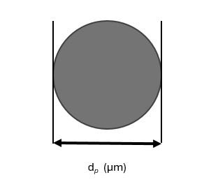
Optical particle counter (OPC) - area-equivalent circle
Particle measurement in the OPC is often based on the principle of light blockage. The size of the particles is determined by the area of the cast shadow and calculated as the diameter of the area-equivalent circle (dp) of this shadow in µm. The number of particles is determined by the number of counting events.
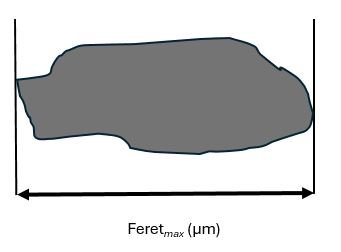
Microscopic evaluation - particle length
Particle measurement under the microscope is carried out by manually or automatically counting and measuring the particles. The size of the particles is determined by the particle length (Feretmax), measured in µm as the largest possible perpendicular distance between two parallels touching the particle. The number of particles is determined by the number of visible particles on the filter.
How comparable are the analysis results?
The analysis results between optical particle counters and microscopic evaluation are not comparable. CleanControlling has dealt intensively with this issue and assesses the comparability of the analysis methods on the basis of the following observations:
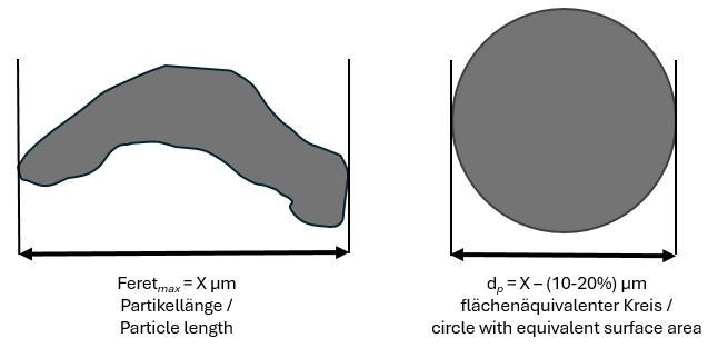
Two fundamental differences often lead to fundamentally different results. Firstly, the value of the area-equivalent diameter is always smaller than the particle length according to Feretmax. The only exception is the circle or sphere. Here Feretmax = dp applies. Furthermore, the influence of the orientation of the particle relative to the “light source - sensor” axis at the moment of detection must be taken into account. In the microscopic analysis, since the particle rests on a filter, it can be assumed that it presents itself with the largest contact surface. As a first approximation, the measured length therefore corresponds to the true Feretmax value.
The examination using OPC, on the other hand, takes place in a moving fluid (gas or liquid). The probability is therefore high that, at the moment of detection, the particle is in a random orientation relative to the light axis and does not present itself with the largest surface area. Whether the shadow cast in this orientation and thus dp is larger or smaller than the value obtained by light microscopic examination depends very much on the geometry of the particle. Since each particle is only photographed once and in a random orientation, the value for dp determined in this way can deviate considerably from the value obtained by light microscopy.
Simulations have shown that convex particles (without recesses) have on average 0-20% higher values for dp when measured using OPC. Concave particles (with recesses, see example) show on average a 10-25% smaller value for dp.
Relevant standards
The following standards are mainly relevant for testing the particle load of medical devices or their cleaning processes:
USP 788
USP 788 describes the requirements and methods for determining the number of particles in injections and parenteral infusions. Two main methods are described: light blocking (OPZ) and microscopic counting.
VDI 2083 Sheet 21
This guideline provides a risk-based approach to the identification and assessment of particle contamination in medical devices during the manufacturing process. It specifies acceptance criteria and methods for checking cleanliness.
VDA 19.1 / ISO 16232:2018
VDA 19.1 or ISO 16232:2018 is the guideline for testing the technical cleanliness of automotive components, which can and is also applied to medical devices. It describes procedures for particle analysis and specifies the classification of particle sizes. The standard test specified here is automated light microscopic analysis with measurement (Feretmax), counting and typing according to metallic shiny and non-shiny.
Conclusion
Both methods, the optical particle counter and microscopic evaluation, have their specific advantages and disadvantages. The choice of method depends on the type of sample and the specific requirements of the respective standard.
Optical particle counters are more suitable for fast and automated solutions, while microscopic analysis provides detailed information about the particle composition. Both methods have specific areas of application and can complement each other to ensure comprehensive particle analysis.
Newsletter registration